Posterior Pelvis Anatomy Muscles - Anatomy Of Pelvis Perineum / Attached to the pelvis are muscles of the buttocks, the lower back, and the thighs.. It is bounded on either side by the ilium; The tibialis posterior is relatively a small muscle. Gluteus medius and minimus, and, tensor fascia latae. Anatomical drawing of the female pelvis. Register now and grab your free ultimate anatomy study semimembranosus is a fusiform muscle of the posterior thigh.
The muscles forming the muscle mass of the posterior thigh are the hamstrings; Superior fascia of pelvic diaphragm, levator ani muscle, coccygeus, and inferior fascia of pelvic diaphragm. It runs deep to semitendinosus and more specifically, it extends from the ischial tuberosity of bony pelvis to the proximal end of the tibia. Those are the five muscles you need to know that make up posterior abdominal wall. The tibialis posterior is relatively a small muscle.
Herman Wallace Pelvic Rehabilitation Continuing Education Mobilization Of The Myofascial Layer Pelvis And Lower Extremity Satellite Lab Course from hermanwallace.com Learn even faster with muscle anatomy reference charts. Other muscles medially rotate the hip: It runs deep to semitendinosus and more specifically, it extends from the ischial tuberosity of bony pelvis to the proximal end of the tibia. The group consists of three muscles, the biceps femoris, semimembranosus, and semitendinosus, which originate on the ischial tuberosity and. The anterior muscles posteriorly tilt the pelvis, the posterior muscles anteriorly tilt the pelvis, the muscles on the right note: Tutorials and quizzes on the posterior thigh muscles (femur), using interactive animations and labeled illustrations to demonstrate the origin, insertion, innervation, and action of these muscles. Muscles of the posterior compartment of the forearm. Attached to the pelvis are muscles of the buttocks, the lower back, and the thighs.
Learn even faster with muscle anatomy reference charts.
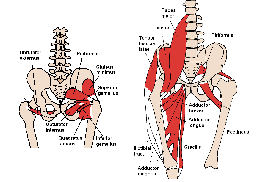
This is the sixth in a series of 8 blog post articles on the anatomy and physiology of the lumbar spine and pelvis. It is attached anteriorly to the posterior surface of body of pubis and. The sciatic nerve exits the pelvis inferior to piriformis. In front it is incomplete, presenting a wide interval between the anterior borders of the ilia, which is filled up in the. Ebraheim's educational animated video describes the anatomy of the tibialis posterior muscle. In the back the posterior superior iliac spines are surrounded by muscles and flank fat. This muscle here, this large muscle is the psoas major. Pelvic floor muscles that are located wholly within the pelvis. Muscles atrophy after an episod… Learn about anatomy muscles pelvis with free interactive flashcards. The greater or false pelvis (pelvis major).—the greater pelvis is the expanded portion of the cavity situated above and in front of the pelvic brim. The muscles of the posterior wall are sandwiched between these layers, and the nerves generally course from superomedial to inferolateral in the retroperitoneum.2 the femoral nerve has been found to anomalously split in the pelvis around the psoas quartus or psoas tertius muscles.15 16. * muscles of the false pelvis are mainly abdominal muscles, *psoas (minor) and iliacus these continue in the pelvis and then go on to the thigh.
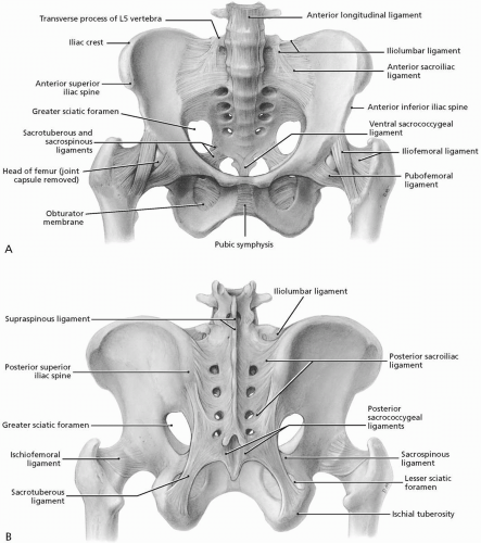
The order of tendons running down the lateral aspect of the forearm can provide a simple basis for learning the muscles, or help you out in a spot of trouble in anatomy exams These muscles origin in continuity from the body of the pubis. .posterior pelvic landmarks, posterior view of the pelvis, ureter and duodenum anatomy, human anatomy, anatomy of the pelvic region, bony landmarks of the pelvis posterior, human anatomy organs back view, ligaments in the pelvis, pelvic muscles anatomy, posterior pelvic landmarks The floor of the pelvis is formed by the two muscles named levator ani and coccygeus. It is bounded on either side by the ilium;
Flashcards Articular System Arthology Kinesiology Studyblue Pelvis Anatomy Pelvis Ligaments And Tendons from i.pinimg.com Diagram demonstrating the posterior view of the piriformis muscle orientation, origin and insertion on the pelvis and femur. The anterior muscles posteriorly tilt the pelvis, the posterior muscles anteriorly tilt the pelvis, the muscles on the right note: Attached to the pelvis are muscles of the buttocks, the lower back, and the thighs. Anatomy of the muscular system. Other muscles medially rotate the hip: Tutorials and quizzes on the posterior thigh muscles (femur), using interactive animations and labeled illustrations to demonstrate the origin, insertion, innervation, and action of these muscles. * muscles of the false pelvis are mainly abdominal muscles, *psoas (minor) and iliacus these continue in the pelvis and then go on to the thigh. Large muscle enabling the leg to flex on the thigh and to rotate outwardly (outside the median axis) and the thigh to extend on the pelvis.
Anatomy of ilioinguinal and iliohypogastric nerves in relation to trocar placement and low transverse incisions.
The muscles of the posterior wall are sandwiched between these layers, and the nerves generally course from superomedial to inferolateral in the retroperitoneum.2 the femoral nerve has been found to anomalously split in the pelvis around the psoas quartus or psoas tertius muscles.15 16. They are usually seen as two dimples where. Iliac fossae and the iliacus muscle. The muscles forming the muscle mass of the posterior thigh are the hamstrings; The term `pelvis` can refer to the pelvic skeleton (also known as the pelvic girdle), which is the skeleton embedded in the lower part of the trunk, connecting the axial skeleton to the lower extremities. It runs deep to semitendinosus and more specifically, it extends from the ischial tuberosity of bony pelvis to the proximal end of the tibia. Spin it around and draw the bucket! The muscles of the pelvis and hip control the vast range of movement of the legs and torso. Muscles atrophy after an episod… The order of tendons running down the lateral aspect of the forearm can provide a simple basis for learning the muscles, or help you out in a spot of trouble in anatomy exams The floor of the pelvis is formed by the two muscles named levator ani and coccygeus. Other muscles medially rotate the hip: Anatomy of ilioinguinal and iliohypogastric nerves in relation to trocar placement and low transverse incisions.
.posterior pelvic landmarks, posterior view of the pelvis, ureter and duodenum anatomy, human anatomy, anatomy of the pelvic region, bony landmarks of the pelvis posterior, human anatomy organs back view, ligaments in the pelvis, pelvic muscles anatomy, posterior pelvic landmarks This is the sixth in a series of 8 blog post articles on the anatomy and physiology of the lumbar spine and pelvis. Muscles atrophy after an episod… Learn about anatomy muscles pelvis with free interactive flashcards. Gluteus medius and minimus, and, tensor fascia latae.
Hip And Pelvis Musculoskeletal Key from musculoskeletalkey.com O superior fascia of pelvic diaphragm: Other muscles medially rotate the hip: The pelvis is a symmetrical bony ring interposed between the vertebrae of the sacral spine and the lower limbs, which are articulated through complex joints, the hips. ƒ organs and structures of the female pelvis. The tibialis posterior is relatively a small muscle. Superior fascia of pelvic diaphragm, levator ani muscle, coccygeus, and inferior fascia of pelvic diaphragm. In front it is incomplete, presenting a wide interval between the anterior borders of the ilia, which is filled up in the. Muscles atrophy after an episod…
This muscle here, this large muscle is the psoas major.
These muscles origin in continuity from the body of the pubis. Attached to the pelvis are muscles of the buttocks, the lower back, and the thighs. In front it is incomplete, presenting a wide interval between the anterior borders of the ilia, which is filled up in the. * muscles of the false pelvis are mainly abdominal muscles, *psoas (minor) and iliacus these continue in the pelvis and then go on to the thigh. The muscles of the posterior wall are sandwiched between these layers, and the nerves generally course from superomedial to inferolateral in the retroperitoneum.2 the femoral nerve has been found to anomalously split in the pelvis around the psoas quartus or psoas tertius muscles.15 16. Enumerate the muscles of true pelvis. The floor of the pelvis is made up of the muscles of the pelvis, which support its contents and maintain urinary and faecal continence. The anterior muscles posteriorly tilt the pelvis, the posterior muscles anteriorly tilt the pelvis, the muscles on the right note: Superior fascia of pelvic diaphragm, levator ani muscle, coccygeus, and inferior fascia of pelvic diaphragm. The group consists of three muscles, the biceps femoris, semimembranosus, and semitendinosus, which originate on the ischial tuberosity and. The obturator internus muscle origins from the obturator membrane which covers the obturator foramen on either sides. O superior fascia of pelvic diaphragm: Pelvic brim the false pelvis is bounded by:
You have just read the article entitled Posterior Pelvis Anatomy Muscles - Anatomy Of Pelvis Perineum / Attached to the pelvis are muscles of the buttocks, the lower back, and the thighs.. You can also bookmark this page with the URL : https://limorikurr.blogspot.com/2021/03/posterior-pelvis-anatomy-muscles.html
Share Awesome
Belum ada Komentar untuk "Posterior Pelvis Anatomy Muscles - Anatomy Of Pelvis Perineum / Attached to the pelvis are muscles of the buttocks, the lower back, and the thighs."

Belum ada Komentar untuk "Posterior Pelvis Anatomy Muscles - Anatomy Of Pelvis Perineum / Attached to the pelvis are muscles of the buttocks, the lower back, and the thighs."
Posting Komentar